



OXIDATIVE STRESS, ASSOCIATED DISEASES, AND LABORATORY EVALUATION
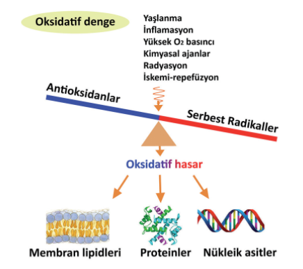
“Under normal physiological conditions, there is a balance between the free radicals and reactive oxygen species (ROS) that are constantly produced in cells and the antioxidants that interact with them. A disruption of this balance in favor of free radicals and ROS—that is, the accumulation of radicals such as superoxide within the cell or the failure of endogenous defense systems—is defined as oxidative stress.”
Free Radicals
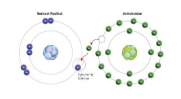
These are compounds that contain unpaired electrons in one of their outer orbitals. They are reactive and short-lived.
Free radicals can be produced as a result of normal metabolic processes or through many reactions necessary for energy production within the cell.
It is generally accepted that free radicals are formed in three main ways.
1- The homolytic cleavage of a normal covalently bonded molecule, in which one of the shared electrons remains in each fragment.
X : Y → X• + Y•
2- When a normal molecule loses an electron
A – e- → A.+ e
3- The addition of a single electron to a normal molecule
A + e- → A•-
It is well known that these radicals, once produced, affect membrane lipids, intracellular proteins, and nucleic acids, leading to changes in the structure and function of these macromolecules and causing cellular damage.
Reactive Oxygen Species (ROS)
Oxygen in the atmosphere is called molecular oxygen (O₂) or dioxygen. A small portion of normal oxygen is reduced during metabolism in cellular compartments—primarily the mitochondria—and converted into reactive oxygen species. The main reactive oxygen species are the superoxide radical (O₂⋅⁻), the hydroxyl radical (OH•), and hydrogen peroxide (H₂O₂). The first two are free radicals, while hydrogen peroxide is a prooxidant.
Antioxidants
Substances that can prevent or delay the oxidation of substances in cells—such as lipids, proteins, and DNA—by free radicals are called antioxidants, and the mechanisms by which they function are referred to as antioxidant defense systems. Antioxidants prevent cellular damage by transferring electrons to free radicals.
Antioxidants have four different mechanisms:
(1) The cleansing effect involves neutralizing free oxygen radicals by either trapping them or converting oxidants into weaker molecules. Antioxidant enzymes and micromolecules work in this way.
(2) Antioxidant effect: The process of neutralizing oxidants by transferring a hydrogen atom to them, or reducing their effects or reaction rates. Vitamins and flavonoids exert their effects in this way.
(3) Repair effect: the repair of biological damage caused by free radicals to structures such as lipids, proteins, and DNA.
(4) Chain-breaking effect: binding free oxygen radicals, breaking their chains, and inhibiting their activity.
Antioxidants also remove oxygen from the environment or reduce its concentration locally, and they remove catalytic metal ions from the environment. These mechanisms can also be included in their mechanisms of action.
Antioxidants;
They are classified into two groups: endogenous (antioxidant enzymes, etc.) and
exogenous (vitamins, etc.).
Table 1. Exogenous Antioxidants
Antioxidant – Mechanism of Action
Vitamin C (Ascorbic acid) – Scavenges hydroxyl radicals (OH•).
Vitamin A (β-Carotene) – Scavenges fat-soluble radicals and singlet oxygen.
Vitamin E – Fat-soluble, exhibits chain-breaking activity.
Phenolic compounds—which are commonly found in vegetables, fruits, spices, and grains—as well as flavonoids such as resveratrol, quercetin, and catechin, along with selenium and zinc, are other important exogenous antioxidants.
Table 2. Endogenous Antioxidants
Non-Enzymatic Compounds
, Albumin—Binds to copper and heme groups, removes HOCl from the environment.
Seruloplasmin – Binds copper ions and uses H₂O₂ to remove copper facilitates its reoxidation.
Transferrin – Binds iron ions in the ferric state (Fe³⁺)
Lactoferrin – Binds ferric iron ions (Fe³⁺) at low pH levels .
Haptoglobin – Binds to hemoglobin.
Hemopexin – Binds to the heme group.
Bilirubin – Scavenges peroxyl radicals.
Glucose – Scavenges hydroxyl radicals (OH•).
Urate – Clears free radicals and binds metals.
Melatonin – Neutralizes hydroxyl radicals (OH•).
Mucus – Removes hydroxyl radicals (OH•).
Enzymes
, Superoxide Dismutase (SOD)
It scavenges the superoxide radical (O2-)
O2- + 2H+ → H2O2 + O2
Catalase (CAT)
It removes hydrogen peroxide (if present in high concentrations) from the environment.
2H₂O₂ + O₂ → 2H₂O + O₂
Glutathione peroxidase (GPx)
It removes hydrogen peroxide (if present at low concentrations) from the environment.
H₂O₂ + 2 GSH → GSSG + 2H₂O
Cytochrome Oxidase
By preventing the release of reactive oxygen species into the environment during the reduction of oxygen in water, it inhibits the formation of ROS (H₂O₂, OH•, O₂⁻).
GSH: Reduced glutathione, GSSG: Oxidized glutathione
Diseases Associated with Oxidative Stress
Reactive oxygen species formed as a result of increased oxidative stress attack the double-bonded groups in intracellular lipids and proteins, as well as the double bonds in DNA bases, and trigger chain oxidation reactions by removing a hydrogen atom. As a result, macromolecules such as intracellular lipids, proteins, and DNA are damaged, leading to cellular damage or cell death.
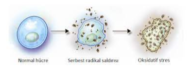
Oxidative stress triggered by free radicals:
1. Neurological disorders (such as Parkinson’s, Alzheimer’s, Huntington’s, amyotrophic lateral sclerosis, multiple sclerosis, depression, and memory loss),
2. Immune system disorders,
3. Diabetes,
4. Rheumatoid arthritis,
5. Kidney diseases (glomerulonephritis, tubulointerstitial nephritis, renal failure, proteinuria, and uremia),
6. Cardiovascular disorders (atherosclerosis, ischemia, hypertension, cardiomyopathy, cardiac hypertrophy, and congestive heart failure, etc.),
7. Chronic obstructive pulmonary diseases,
8. Ischemia-reperfusion injury,
9. Inflammatory diseases,
10. It is believed to contribute to the development of over a hundred diseases, including cancer.
Furthermore, the progressive nature of oxidative damage caused by free radicals contributes to aging and the onset of age-related degenerative diseases (such as cataracts and atherosclerosis).
Laboratory Tests Used to Assess Oxidative Stress
The reactivity and short half-lives of free radicals are a major factor preventing the direct measurement of these substances. For this reason, many of the methods developed to measure these substances and their products in various tissues and materials are indirect and do not directly measure free radicals.
Lipid peroxidation occurs through a chain reaction in which unsaturated fatty acids in membrane phospholipids react with oxygen to form lipid hydroperoxides (LOOH). As a result of a series of reactions, MDA (malondialdehyde), certain other aldehydes, conjugated dienes, and volatile Products such as hydrocarbons are released. By identifying one of these released intermediate products, lipid peroxidation and, indirectly, free radicals are measured.
By measuring the levels of the antioxidant enzymes Superoxide Dismutase (SOD) and Glutathione Peroxidase (GPx), one can gain insight into these components of the antioxidant system.
Antioxidant vitamins—such as Vitamin A, Vitamin C, and Vitamin E—can be measured using methods like spectrophotometry or the more sensitive HPLC, while extracellular antioxidants such as uric acid, bilirubin, and albumin can be determined using routine colorimetric methods. Measurements of plasma selenium, intracellular zinc in erythrocytes, and plasma levels of ceruloplasmin, haptoglobin, transferrin, and hemopexin are also valuable in assessing antioxidant capacity.
In addition, by measuring Total Antioxidant Capacity using spectrophotometric methods, a general idea of an individual’s antioxidant capacity can be obtained.
References:
1. Aslankoç R, Demirci D, İnan Ü, Yıldız M, Öztürk A, Çetin M, Savran EŞ, Yılmaz B. The Role of Antioxidant Enzymes in Oxidative Stress—Superoxide Dismutase (SOD), Catalase (Cat), and Glutathione Peroxidase (Gpx). Med J SDU 2019; 26(3): 362-369.
2. Tanakol A, Uzunçakmak TK, Kutlubay Z. Oxidative Stress and Aging. Dermatoz 2020;11(3):31-35.
3. Özcan O, Erdal H, Çakırca G, Yönden Z. Oxidative Stress and Its Effects on Intracellular Structures. JClin Exp Invest 2015; 6 (3): 331-336.
4. Dündar Y. Oxidative Stress and Antioxidants in Medicine. ISBN: 9789757150299. Afyon Kocatepe University; Pub. No. 29. 2000.