
Egelab

Neurological Symptoms of Unexplained Cause In patients
This diagnosis should bring this to mind!

PARANEOPLASTIC NEUROLOGICAL SYNDROMES

Paraneoplastic Neurological Syndrome (PNS) is a condition that occurs in patients with cancer and is not caused by the direct or local effects of the underlying tumor , nor by metastases, opportunistic infections, or side effects of cancer treatment , and for which a significant proportion is believed to result from autoimmune mechanisms .
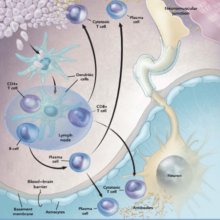
Paraneoplastic Neurological Syndrome (PNS) is a condition that occurs in cancer patients and is not caused by the direct or local effects of the underlying tumor , nor by metastases, opportunistic infections, or side effects of cancer treatment , and for which a significant proportion is believed to result from autoimmune mechanisms .
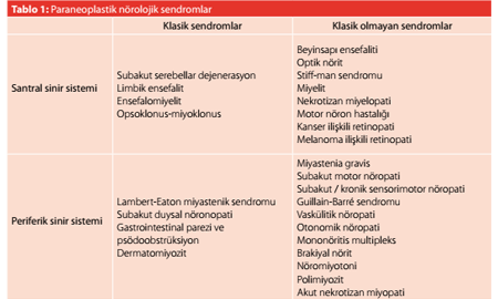
Although it is known that PNSs are frequently observed (3–15%) in the course of small cell lung cancer (SCLC), thymoma, monoclonal gammopathy, and hematologic malignancies , regarding the prevalence of PNS in all cancer cases, various studies have reported values ranging from 1/1,000 to 1/10,000 . Although PNSs occur rarely , they are significant because in most cases they develop before a cancer diagnosis is made and generally when the cancer is still very small and treatable . PNS findings that emerge following a cancer diagnosis may mimic neurological syndromes associated with opportunistic infections ( ) and the side effects of cancer treatment ( ). Furthermore, syndromes frequently observed in cancer patients—such as limbic encephalitis or subacute cerebellar degeneration —do not always indicate an underlying cancer . Some neurological syndromes are more commonly seen in association with cancer or their clinical features directly suggest a paraneoplastic etiology. These are referred to as “classic” PNS. Some syndromes, however, typically occur in patients without cancer. These are also referred to as “non-classical” syndromes. In “non-classic” syndromes such as brainstem encephalitis, myasthenia gravis, and polymyositis , the likelihood of finding an associated cancer is low, and in these cases, it is necessary to prioritize screening for other complications of cancer . In addition, “classic syndromes” associated with antibodies directed against ion channels can often present without an accompanying tumor. For example, in cases of limbic encephalitis where voltage- -potassium channel (VGKC) antibodies are detected , a tumor has been identified in only 20% of cases.
The most widely accepted view today is that the underlying tumor… similar antigenic properties between the nervous systems an autoimmune response resulting in the emergence of PNSs He is responsible for its emergence. In recent years, some Serum and cerebrospinal fluid (CSF) of PNS cases synthesized by various tumors in the samples nervous system antigens (paraneoplastic or antibodies developed against onconeural antigens This has been determined. In a significant number of these cases that there is intrathecal onconoral antibody synthesis This demonstrates the role of these antibodies in the pathogenesis of PNSs. This led people to believe that he was playing a role. Also affected nerve The presence of inflammatory infiltration in systemic fields, via passive transfer of some onchoneural autoantibodies to enable the creation of experimental animal models and removes autoantibodies from circulation, such as plasmapheresis treatment methods lead to clinical improvement in some cases The fact that it leads to this has supported this view. In PNS cases identified autoantibodies, central and peripheral nerve in the membranes of system neurons or cells contained within (cytoplasm and/or nucleus) It develops in response to antigens. The diagnosis of PNSs involves recognizing the neurological syndrome and its associated components. cancer detection and serum/CSF antibodies It is diagnosed by detection.
Image of an IFA preparation showing autoantibodies directed against neurons.
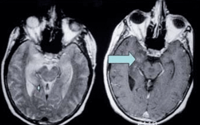
For the detection of nervous system disorders and cancer. Imaging, CSF and EEG studies are used in the diagnostic process. These are methods that could be useful. CSF shows lymphocytic pleocytosis, increased protein, and high IgG levels. index and oligoclonal band presence are frequently encountered. These are the findings. CSF analysis also It should also be done to rule out leptomeningeal metastasis, and presence of atypical cells in the obtained CSF sample It should be investigated. Imaging of the affected area in all PNS cases. MRI scan (especially T2 and FLAIR) should be performed. (weighted sequences) metastasis and other cancer-related Useful for ruling out central nervous system diseases. It is a method. For the same reason, contrast enhancement should also be examined in these studies. Most PNS In this case, the blood-brain barrier (BBB) ​​was preserved and Therefore, the probability of finding a contrast-enhancing lesion. The value is low. EEG examination reveals many paraneoplastic abnormalities. Non-convulsive symptoms that can be seen in encephalitis cases It is important to ensure that the status is not overlooked, and especially in cases presenting with confusion and altered consciousness should be implemented. PNSs usually develop in the early stages of cancer and Therefore, it is often possible to visualize the tumor. It may not be the case. The underlying tumor may be in the breast, abdomen, and pelvis. This can be shown with a CT scan. The syndrome or Identification and visualization of paraneoplastic antibodies. It enables better selection of the target area. For example, in a case of anti-Yo-positive cerebellar ataxia, it is important to perform a mammogram and gynecological examination, or in a case of anti-Ma-positive encephalitis, to perform a testicular ultrasound. 18F-Fluorodeoxyglucose (FDG)-PET imaging allows for the detection of small primary tumors and metastases and helps identify potential sites for biopsy.
A. MRI image of a patient with cerebellar involvement
B. MRI image of a patient with bilateral temporal lobe involvement

Patients with classic PNS and those who test positive for well-characterized paraneoplastic antibodies but in whom no underlying tumor has been identified should be closely monitored for 5 years. In 80–90% of these patients, an underlying tumor becomes apparent within the first year. Cases in which classic PNS findings develop while the cancer is in remission should also be evaluated for cancer recurrence.

Diagnostic criteria have been proposed for the diagnosis of PNS. According to these criteria, PNS cases are classified into two categories—definite and probable—based on the presence of cancer, well-characterized antibodies, and a classic syndrome:

Definitive PNS criteria:

  1. Diagnosis of the classic syndrome and cancer (within 5 years of the syndrome’s diagnosis).
  2. Identification of a nonclassical syndrome that has resolved or significantly improved following treatment of the tumor (there should be no concomitant immunotherapy, and the syndrome should not be a condition that can resolve spontaneously, such as myasthenia gravis).
  3. Detection of antineuronal antibodies in association with a non-classical syndrome and cancer (with an interval of no more than 5 years).
  4. The detection of well-characterized paraneoplastic antibodies (Hu, Yo, CV2, Ri, Ma2, amphiphysin) in the presence of a neurological syndrome (classic or otherwise), even in the absence of a concomitant cancer.

Possible PNS criteria:

  1. The presence of a classic neurological syndrome and a high risk of cancer despite the absence of antibodies or evidence of cancer.
  2. Detection of partially characterized antineuronal antibodies despite the absence of a neurological syndrome (classic or otherwise) or cancer.
  3. Absence of antineuronal antibodies despite the diagnosis of a non-classical syndrome and cancer (within 2 years of the syndrome’s diagnosis).

For the effective treatment of PNS, it is important to recognize the clinical syndrome early, identify the underlying tumor and associated antibodies, and rapidly determine the paraneoplastic etiology. Once the diagnosis of PNS is confirmed, treatment proceeds to the stages of effective tumor therapy and immunosuppressive therapy. In cases where a tumor is detected, treatment should be initiated with tumor resection and chemotherapy, along with steroids, IV Ig, or plasmapheresis.

While the response to treatment is poor in syndromes associated with intracellular antigens—even after tumor treatment and immunosuppression—the response to treatment is good in syndromes associated with cell membrane antigens. Even in this second group, patients may die if treatment is not started early and the underlying tumor is not removed. For this reason, early diagnosis and treatment are the most important factors affecting the prognosis of PNSs.

REFERENCE:

Erdem TÜZÜN. Paraneoplastic Syndromes with Neurological Manifestations. Clinical Development. 2010-1, pp. 71–77.

Darnell, R.B. et al. Mechanisms of Disease: Paraneoplastic Syndromes Involving the Nervous System. N Engl J Med 2003;349:1543-54.

Leave a Reply

Your email address will not be published. Required fields are marked *